Great How To Understand Ultrasound Scan Report

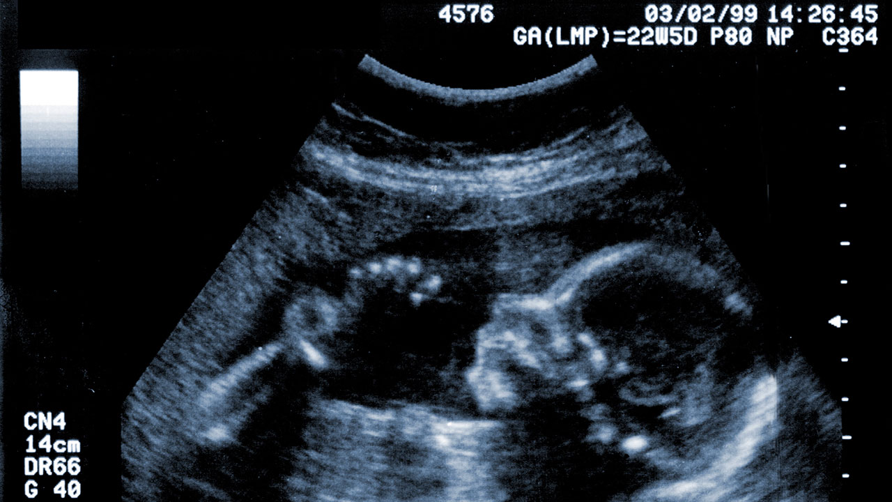
The morphology scan is a detailed ultrasound scan that looks at your babys body and observes the posit.
How to understand ultrasound scan report. Hello The ultrasound report shows the presence of an abdominal mass measuring 1 cm in all three dimensions. First for the purposes of ophthalmic ultrasound the posterior of the eye is centered on the optic nerve not the fovea. The mass needs to be removed surgically since it could be a cyst arising from ovary.
It could also show up certain defects for instance a cleft lip or palate. Fed up with deciphering jargon Dr Attiya Khan asked consultant gynaecologist Mr Rehan Khan for a plain language guide to understanding pelvic ultrasounds. Anomaly pregnancy ultrasound what is anomaly scan.
The process sends high-frequency low-power waves to the uterus in the womans stomach and these waves bounce back when they hit the surface of the fetus and detect changes. A computer is used. The simple truth is that radiology reports can be hard to read especially for those without a medical background.
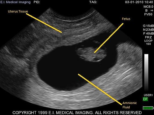
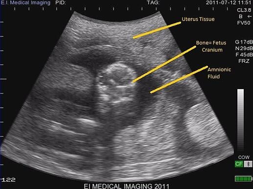
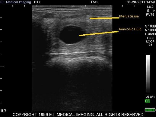
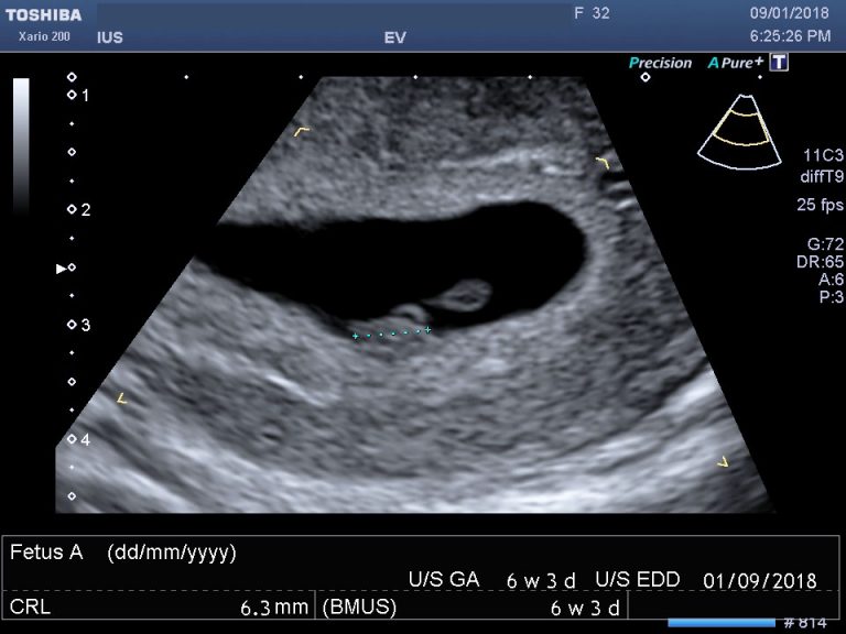
13 22 23 25 29 - 31 In general an ultrasound report should contain the following sections. An ultrasound or sonogram picture is a black and white photograph so they all look the same to someone who doesnt know much about how to read an ultrasound. The combination of advanced medical technology and the wonderful subtle intricacies of the human body often result in a final document that more closely resembles a William.
Fatty change is when fat builds up in your liver cells. The CIMT Test report provides quite a bit of information and it is broken down in to sections. An insider guide to reading your radiology report.
The mass is septate and there is calcification in the wall. We will cover different types of ultrasound scans in a future section of this post but for now here is a quick summary of what a 3D and 4D scan will offer. A 3D ultrasound scan will be able to show you some of the features on your babys face.
